What Is Adenomyosis? This Little-Known Condition Affects Up to 1 in 5 Women
A widespread but seldom-discussed disorder is causing severe reproductive and health problems, highlighting a major oversight in women's healthcare.
STORY AT-A-GLANCE
Adenomyosis is a benign estrogen-dependent uterine disorder that may cause abnormal uterine bleeding, pelvic pain and infertility
It’s estimated that about 20% of women, primarily those of reproductive age, may suffer from the condition, yet there’s surprisingly little awareness surrounding it
Estrogen is believed to promote the growth of adenomyosis; many people are exposed to excessive estrogen in the form of birth control, estrogen replacement therapy and even exposure to plastics
Estrogen is also carcinogenic and antimetabolic, radically reducing the ability of your mitochondria to create cellular energy
One of the most important strategies for adenomyosis — aside from avoiding estrogen and xenoestrogens — is to take natural progesterone, which is anti-estrogenic
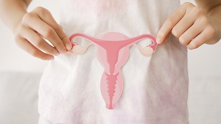
Adenomyosis is a benign estrogen-dependent uterine disorder,1 which involves symptoms such as abnormal uterine bleeding, pelvic pain and infertility.2 In cases of adenomyosis, endometrial tissue, which normally lines the uterus, is found in the myometrium, or the muscular wall of the uterus.
In other words, the uterine lining grows into the uterus' muscular wall,3 causing symptoms ranging from mild to severe. For some women, adenomyosis causes no symptoms at all while others experience debilitating pain.
It's estimated that about 20% of women, primarily those of reproductive age, may suffer from the condition,4 yet there's surprisingly little awareness surrounding it among health care professionals and the public alike.5
What Are the Signs and Symptoms of Adenomyosis?
Adenomyosis can be categorized into two main types: focal and diffuse. In focal adenomyosis, the endometrial tissue is localized to a specific area or areas within the uterine muscle. These localized regions are sometimes referred to as adenomyomas, which are similar to fibroids but contain endometrial tissue.
Diffuse adenomyosis involves a more widespread infiltration of endometrial tissue throughout the uterine muscle. This type is characterized by a uniformly thickened uterine wall. In either type, the lining of the uterus grows into the uterine wall, leading to thickening and enlargement of the uterus, which in some cases may double or triple in size.6
"The displaced tissue continues to act normally — thickening, breaking down and bleeding — during each menstrual cycle," Mayo Clinic notes.7 Common symptoms of adenomyosis include heavy menstrual bleeding, severe menstrual cramps, pelvic pain and bloating.
Some women with adenomyosis may also experience pain during intercourse. Infertility is also possible and women with adenomyosis who become pregnant are more likely to have complications such as miscarriage, pre-term delivery, preeclampsia and post-delivery bleeding.8
The condition can sometimes be asymptomatic, however, making diagnosis challenging. While most women who develop adenomyosis are between the ages of 40 and 50, it can develop in younger women as well. The condition is becoming more common in women in their 30s,9 and even teenagers can be affected. Other factors that increase the risk of adenomyosis include:
Carrying two or more pregnancies to term10
Prior uterine surgeries, including uterine fibroid removal, cesarean section or dilatation and curettage (D&C)
Endometriosis
Top Causes of Adenomyosis
A combination of hormones, genetics and inflammation or injury to the area likely influence adenomyosis development. Science Alert reported:11
"It is thought that the region between the endometrium and myometrium becomes damaged, either by the natural processes of the menstrual cycle, pregnancies, and childbirth, or medical procedures. In some women, damage to the endometrial tissue layer does not heal as it should, and the endometrium-like cells enter and grow abnormally into the myometrium."
It's possible that uterine incisions during surgery may allow for the direct invasion of endometrial cells into the uterine wall, for instance. It's also been suggested that the misplaced endometrial tissue may be deposited in the uterine wall in the fetus during development.12
Inflammation of the uterine lining after childbirth is another potential factor that could degrade the boundary of cells lining the uterus. Another theory suggests bone marrow stem cells in uterine muscle could trigger the condition.13
Estrogen, a hormone involved in the menstrual cycle, is also believed to promote the growth of adenomyosis. Therefore, conditions that result in higher levels of estrogen may be linked to an increased risk. "Regardless of how adenomyosis develops, its growth depends on the body's circulating estrogen," Mayo Clinic explains.14
Estrogen Promotes Adenomyosis
"Estrogens have been proven to be the crucial hormones driving the growth of adenomyosis," as noted in the Journal of Clinical Medicine.15 Yet, estrogen's exact role in worsening adenomyosis is still not fully understood.
In one study, researchers found that a molecule called miR-145 is much higher in adenomyosis tissue compared to healthy tissue.16 High levels of miR-145 turn on genes that cause inflammation and cell adhesion.
One important gene affected by miR-145 is CITED2, which plays a key role in stopping cell movement. The study showed that estrogen, through a specific receptor, increases miR-145 levels, helping adenomyosis to progress. Yale Medicine also explains:17
"Evidence suggests that prolonged exposure to estrogen may be a contributing factor [to uterine adenomyosis]. Women may be exposed to more estrogen if their menstrual cycles are shorter than average or if they have been pregnant two or more times."
However, many people are exposed to excessive estrogen in the form of birth control, estrogen replacement therapy and even exposure to plastics.
The Hazards of High Estrogen
While estrogen is routinely referred to as "the female hormone," this is misleading, as it's not exclusive to women. Moreover, there's not just one estrogen but several. One of the properties of estrogens is their ability to increase cells' ability to hold water, which is why women with estrogen dominance are prone to edema (water retention). Cellular swelling is both a characteristic of the cellular stress response and a signal for cellular proliferation.
During the follicular phase of the menstrual cycle, estrogen stimulates the uterine lining and follicles to swell and multiply in preparation for the fertilization of an egg. Similarly, during and after pregnancy, breast tissue swells and grows larger to facilitate milk production. But cellular swelling and proliferation is also a hallmark of cancer. Indeed, the word oncology comes from the Greek word "oncos," which means swelling.
In his 1997 book, "From PMS to Menopause: Female Hormones in Context,"18 biologist Ray Peat19 stated that estrogen had been shown to replicate the shock phase of the stress reaction in animals. According to Peat, the physiological purpose of estrogens is to stimulate cell division by triggering water uptake by the cell. Peat also suspected that estrogen was a metabolic inhibitor that slows down energy production in the cell.
Further, as bioenergetic researcher Georgi Dinkov explained in my interview with him, estrogen is carcinogenic and antimetabolic, radically reducing the ability of your mitochondria to create cellular energy in a form of ATP by depending on aerobic glycolysis (the Warburg effect), which radically impairs oxidative phosphorylation. This further contributes to its carcinogenic effect.
According to Dinkov, the biochemical role of estrogen is to aid in wound healing. In cases of tissue trauma, estrogen reverts the differentiated cells in that specific tissue back to a stem cell-like condition, to repair the damaged tissue. In young, healthy women, progesterone will turn off estrogen's activity. However, progesterone declines with age, but estrogen synthesis typically does not. Hence, if your estrogen is high and progesterone low, your cancer risk will rise.
Estrogen also promotes the differentiation and proliferation of fat cells (adipocytes) in the subcutaneous fat layer, which is particularly prevalent in the thighs, hips and buttocks and therefore leads to increased fat storage in these areas.
Some people and clinicians believe that bioidentical estrogen solves the problem but it does nothing of the sort. Bioidentical estrogen still has all the same negative characteristics. It increases the risk of all cancers, lowers your metabolic rate and increases your risk of obesity, diabetes and osteoporosis — along with adenomyosis.
Tips to Lower Your Estrogen Exposure and Load
Conventional adenomyosis treatment typically involves pain medications and hormonal treatments, such as birth control pills, and even surgery, including hysterectomy. But one of the most important strategies — aside from avoiding estrogen and xenoestrogens — is to take natural progesterone. Not only is progesterone anti-estrogenic but it's a potent cortisol blocker and will improve mitochondria production of cellular energy by blocking estrogen and cortisol.
Before you consider using progesterone it is important to understand that it is not a magic bullet and you get the most benefit by implementing a Bioenergetic diet approach that allows you to effectively burn glucose as your primary fuel with backing up electrons in your mitochondria that reduces your energy production. My new book coming out shortly about Cellular Health covers this process in great detail.
Once you have dialed in your diet, an effective strategy that can help counteract estrogen excess is to take transmucosal progesterone (not oral or transdermal), which is a natural estrogen antagonist. Progesterone is one of only four hormones I believe many adults can benefit from. (The other three are thyroid hormone T3, DHEA and pregnenolone.)
I do not recommend transdermal progesterone, as your skin expresses high levels of 5-alpha reductase enzyme, which causes a significant portion of the progesterone you're taking to be irreversibly converted primarily into allopregnanolone and cannot be converted back into progesterone.
As a general recommendation, I recommend taking 25 to 50 mg of bioidentical progesterone per day, taken in the evening one hour before bed, as it can also promote sleep. For optimal bioavailability, progesterone needs to be mixed into natural vitamin E. The difference in bioavailability between taking progesterone orally without vitamin E and taking it with vitamin E is 45 minutes versus 48 hours.
You can make your own by dissolving pure USP progesterone powder into one capsule of a high-quality vitamin E, and then rub the mixture on your gums. Fifty milligrams of powdered progesterone is about 1/32 teaspoon.
Do not use synthetic vitamin E (alpha tocopherol acetate — the acetate indicates that it's synthetic). Natural vitamin E will be labeled "d alpha tocopherol." This is the pure D isomer, which is what your body can use.
There are also other vitamin E isomers, and you want the complete spectrum of tocopherols and tocotrienols, specifically the beta, gamma, and delta types, in the effective D isomer. As an example of an ideal vitamin E you can look at the label on our vitamin E in our store. You can use any brand that has a similar label.
If you are a menstruating woman, you should take the progesterone during the luteal phase or the last half of your cycle which can be determined by starting 10 days after the first day of your period and stopping the progesterone when your period starts.
If you are a male or non-menstruating woman you can take the progesterone every day for 4-6 months and then cycle off for one week. The best time of day to take progesterone is 30 minutes before bed, as it has an anti-cortisol function and will increase GABA levels for a good night's sleep.
Please note that when progesterone is used transmucosally on your gums as I advise, the FDA believes that somehow converts it into a drug and prohibits any company from advising that on its label. However, please understand that it is perfectly legal for any physician to prescribe an off-label indication for a drug.
In this case progesterone is a natural hormone and not a drug and is very safe even at high doses. This is unlike synthetic progesterone, called progestins, that are used by drug companies, but frequently, and incorrectly, referred to as progesterone, which are dangerous and should never be used by anyone.
Considering the health risks associated with estrogen excess, here are some additional commonsense strategies that can help you limit your exposure and lower your estrogen load:
Avoid synthetic estrogens — Minimize exposure to synthetic estrogens, such as those found in hormone replacement therapy and oral contraceptives. Consult with a qualified health care professional about alternative treatments and/or contraceptive methods with lower estrogen content.
Avoid linoleic acid (LA) — Omega-6 PUFA like LA functions very similarly to estrogen as they both increase your risk for cancer and decrease metabolic function by suppressing your thyroid. Best to read my comprehensive LA article for more details.
Choose natural products — Opt for natural and organic personal care products, including makeup, skin care, and hair care items, to reduce exposure to synthetic chemicals like parabens and phthalates, which have estrogenic properties.
Limit pesticide exposure — Choose organic produce whenever possible to reduce exposure to pesticides, many of which have estrogenic effects. Washing fruits and vegetables thoroughly can also help remove pesticide residues.
Rethink your household products — Many household cleaning products, laundry detergents and air fresheners contain chemicals with estrogenic properties. Swap them out for natural, nontoxic alternatives or make your own cleaning solutions using vinegar, baking soda and essential oils instead.
Avoid plastic containers — Minimize the use of plastic containers and food packaging, which can leach estrogenic compounds into food and beverages. Instead, opt for glass or stainless steel containers for food storage and water bottles.
Maintain a healthy weight — Aim for a healthy weight and body composition through a balanced diet and regular exercise. Excess body fat, particularly around the thighs, hips, and buttocks, can contribute to higher estrogen levels.
Support liver health — Support liver function, as your liver plays a crucial role in metabolizing and eliminating excess estrogen from your body. Eat a nutrient-rich diet, limit alcohol consumption and consider incorporating liver-supporting herbs and supplements, such as milk thistle or dandelion root.
Promote hormonal balance — Explore natural approaches to promote hormonal balance, such as consuming foods rich in cruciferous vegetables (such as broccoli, cauliflower and kale) and flaxseeds, which contain compounds that help support estrogen metabolism and detoxification.
Reduce stress — Manage stress through relaxation techniques like meditation, deep breathing exercises, yoga or spending time in nature. Chronic stress can disrupt hormone balance, including estrogen levels, so prioritizing stress reduction is essential.
Disclaimer: The entire contents of this website are based upon the opinions of Dr. Mercola, unless otherwise noted. Individual articles are based upon the opinions of the respective author, who retains copyright as marked.
The information on this website is not intended to replace a one-on-one relationship with a qualified health care professional and is not intended as medical advice. It is intended as a sharing of knowledge and information from the research and experience of Dr. Mercola and his community. Dr. Mercola encourages you to make your own health care decisions based upon your research and in partnership with a qualified health care professional. The subscription fee being requested is for access to the articles and information posted on this site, and is not being paid for any individual medical advice.
If you are pregnant, nursing, taking medication, or have a medical condition, consult your health care professional before using products based on this content.


We know that intestinal dysbiosis, defined by an alteration in the composition of the intestinal microbiota and the reduction of its diversity, is associated with intestinal and extraintestinal conditions, such as irritable bowel syndrome, inflammatory bowel disease, celiac disease, colorectal cancer. , metabolic disorders (obesity and type 2 diabetes), Alzheimer's and Parkinson's diseases and other pathologies.
In this study, gut microbiota diversity was significantly lower in patients with adenomyosis. Furthermore, the analysis showed that the composition of the intestinal microbiota differs between patients with adenomyosis and controls. Patients with adenomyosis presented a reduction of the Bifidobacterium genus in the intestinal microbiota compared to the control group. Bifidobacterium is known to have beneficial effects such as the production of short-chain fatty acids (SCFAs), the formation of biological barriers, and the secretion of antimicrobial compounds. Most of the other bacterial taxa depleted in the adenomyosis group are important butyrate producers and some have also been found to be reduced in the inflammatory bowel disease, Crohn's disease. disease, and endometriosis. Butyrate is a SCFA found in the intestinal metabolome that serves as a crucial energy source for enterocytes and stimulates the growth of the intestinal epithelium, repairing and strengthening the intestinal barrier. A decrease in the abundance of taxa responsible for butyrate production can lead to compromised epithelial cell integrity, increased colonic permeability, bacterial infiltration, and local inflammation.
Strobolome has been considered to contribute significantly to adenomyosis and endometriosis by deregulating circulating estrogen levels in women, through gut microbial enzymes involved in its metabolism. In particular, beta-glucuronidase, beta-galactosidase and beta-glucosidase are bacterial hydrolytic enzymes that play a key role in the deconjugation of estrogens leading to an increase in estrogen. Many studies have shown that inflammatory, angiogenic, growth and hormonal factors may play an important role in the development of adenomyosis. As Dr. Mercola reports, prolonged exposure to estrogen may be a contributing factor to adenomyosis. Bacterial taxa are Gram-negative and, therefore, are characterized by having lipopolysaccharide (LPS) as the main component of their outer membrane. LPS functions as a powerful endotoxin and its activation of pattern recognition receptors, such as the toll-like receptor, plays a crucial role in eliciting pro-inflammatory and immune responses, as well as promoting neoangiogenesis and the secretion of growth factors. It is known that all of these mechanisms are involved in the pathogenesis of adenomyosis. The composition of the intestinal, vaginal and endometrial microbiota of patients with adenomyosis is a factor of important evidence in adenomyosis. Probiotics are beneficial for health and, according to studies in humans and animals, they act to eliminate female gynecological disorders such as vaginal infections, polycystic ovary syndrome, adenomyosis and endometriosis.
https://journals.plos.org/plosone/article?id=10.1371/journal.pone.0263283 (2022).--
https://www.ncbi.nlm.nih.gov/pmc/articles/PMC9369051/ (2022).--
https://www.ncbi.nlm.nih.gov/pmc/articles/PMC11168261/ (2024).—
https://scholar.utc.edu/honors-theses/478/ (2024).—
https://www.ncbi.nlm.nih.gov/pmc/articles/PMC10301998/ (2023).—
https://journals.sagepub.com/doi/10.1177/17455057241234524?icid=int.sj-full-text.similar-articles.3 (2024).--